01 Overview
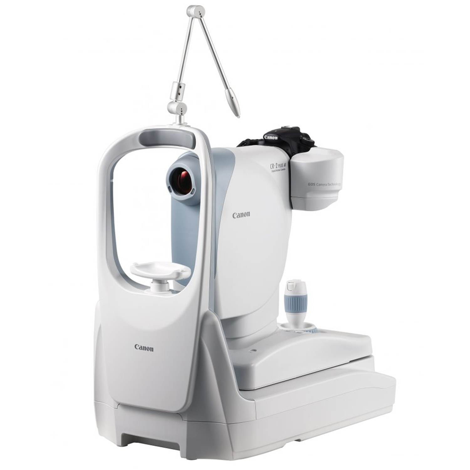
The Canon CR-2 PLUS AF Digital Non-Mydriatic Retinal Camera provides Color and Fundus Autofluorescence (FAF) imaging within a small compact design. Features include Auto-Fundus, Auto-Focus, Auto-Capture and Image Error Detection. Geographic Atrophy, Macular Degeneration, Glaucoma, Diabetic Retinopathy and other conditions that can affect vision may also be identified and monitored using FAF mode. Using invisible infrared alignment light, the digital non-mydriatic camera may image patients with pupils as small as 3.3 mm (small pupil mode) without dilation drops. This is especially useful when performing retinal screenings or expediting routine retinal imaging exams during office visits.


02 Details

Auto-Focus with Manual Alignment Override
Automatically focuses the eye by partially depressing the joystick, or easily switches to manual focus with a twist of the focus ring.
Auto-Capture
Automatically captures the image once the eye is properly focused
Auto-Exposure
Automatically measures the volume of infrared light at the retina and adjusts the flash intensity.
Image Error Detection
Advanced software automatically confirms both correct alignment and focus.
Contrast Enhancement
Provides increased image clarity by emphasizing the difference in ‘redness’ and ‘brightness’ of blood vessel structures relative to their surroundings.

Auto-Fundus
Automatically switches from the external eye to retinal observation mode when the eye is properly aligned.
High Resolution Images

45 degrees (90 degrees measured from the center of the eye)

2X digital magnification. This option can also be used to provide an image with a 30 degrees angle.

With the optional mosaic software up to 20 images can be combined, providing wide field imaging.
The image processing that is applied, reduces the gradation of overexposure. Low-intensity sections (macula) are clearly visible, while maintaining the gradation in tone of the high-intensity portion (the optic disc).

FAF (Fundus Auto Fluorescence)
FAF Imaging for the diagnosis of retinal disease is a relatively new diagnostic technique that provides more information on the health of the retinal pigment epithelium. FAF has proven to be very useful for the early detection of age related Macula Degeneration (AMD), one of the leading causes of visual impairment. Recent studies indicate that FAF Imaging can also aid in the diagnosis of a variety of other diseases and even in the detection of intraocular tumors.

Color

The FAF image shows pathology that can’t be seen in the color image
Digital Filter Processing


With digital red-free, digital cobalt mode, the CR-2 PLUS AF extracts each element from the color image. Images are created from the original RAW information from the digital camera. Based on the EOS retina technology and Canon proprietary image processing, it provides image quality- fully comparable with traditional optical filters.

Quick and easy anterior segment photography to document the cornea, pupil, eyelid and sclera.

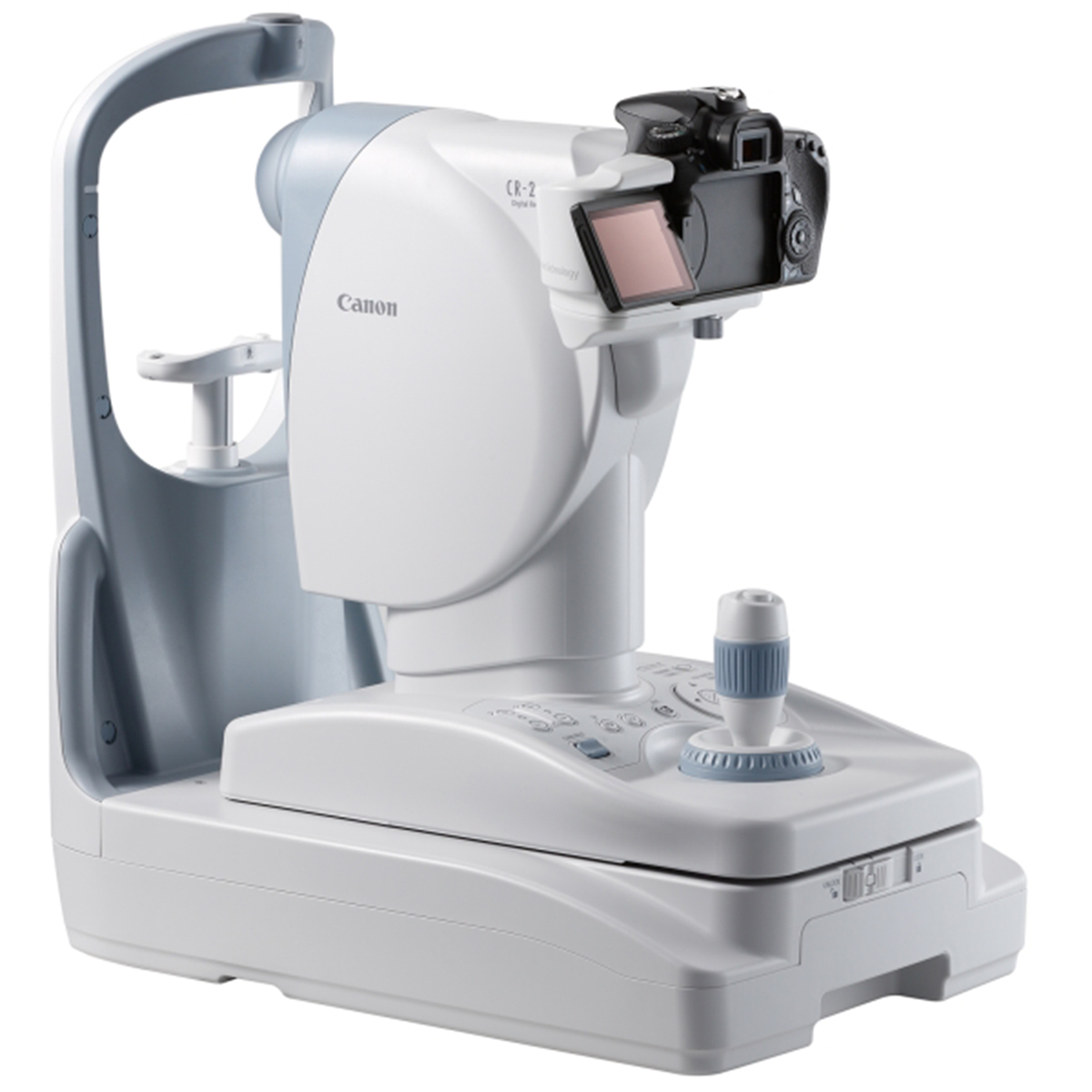
Intelligent design with specifically shaped surfaces to act as grip; intuitive handling for quick and efficient image capture. Switches are illuminated for easy operation in darkened rooms.

Besides the shutter release and aligment buttons, it also includes the up and down movement of optical head (powered) and manual focus control.

The extensive auto functions of the CR-2 PLUS AF will make the examinations much easier. For patients with ocular opacities, involuntary eye movements, lack of co-operation and small pupils the CR-2 PLUS AF also provides full manual control. Manual operation can be crucial to overcome the limitations of fully automatic cameras.

Dedicated 24 Mega Pixel Digital Camera
The EOS Retina camera provides optimal retinal imaging. And it is completely integrated with the functions of the retinal camera. A further advantage is that the digital camera body can be exchanged easily in case of upgrading to a newer model or service requirements.
Full LED Technology
LED technology is used for the observation as well as for the flash, for greater patient comfort, improved reliability and extended life-time.
Low Flash Mode
Ultra low Flash Intensity will reduce patients’ discomfort greatly and essential for photophobic patients. Restriction of pupil is reduced; easy to re-take photos, or to take images of both eyes without waiting.
Retinal Imaging Control Software
Software designed to work with all Canon retinal cameras; for capturing, reviewing and reporting. When the camera is used stand-alone, it also functions as database with an archive function.

Extremely intuitive user interface. Clear graphic representation of functions and display of previous studies.

Displays images of the same side eye to compare them. Or select studies from previous dates to monitor progression.

Observation progression: select up to 5 past examinations to see changes over time.

Report screen with extensive software tools: embossing a retinal image, changing gamma value, brightness and contrast, color balance balance, adding annotations and measuring C/D ratio.
EMBOSS can be useful to enhance the topography of the retina in the evaluation of macular degeneration, glaucoma, and retinopathy.

Emboss Negative
This function will make the blood vessels stand out.

Emboss Positive
The optic disc stands out.

Inversion
Inverts the color of an image to assist diagnosis.

Overlay
Overlay 2 images to see differences and changes in pathology. Selected images are aligned automatically. It can be useful for comparison to see changes, but also the colour image could be overlayed with e.g. a FAF image for additional information.

Annotations
Add shapes; lines, arrows and comments to a captured image.

CUP/DISC Measurement
Measure the optic nerve papillary area. The C/D can be measured as line or area ratio.
Canon Opacity Suppression
When obtaining retinal images, ocular opacity can cause several problems: the scattering of the light will make the edges of the blood vessels appear blurred, the difference in brightness of the retina will be reduced, making it very difficult to distinguish between structures. Furthermore a cataract eye will cause images to appear more yellow. Canon opacity suppression tool is a unique and sophisticated software tool, that based on the full information from the CMOS sensor of the digital camera, will largely suppress the effect of ocular opacities on color images.


Our retinal cameras can be combined with a Canon OCT, using the same software and sharing the database. Output of both devices can be combined in one report to facilitate a comprehensive analysis.
03 Support

Order Enquiry